Anatomy

Among our most widely cited resources, these neuroanatomical atlases provide data-driven, high-resolution maps of the mammalian brain. Each atlas has accompanying software tools for streamlined use by the community.

Adult Mouse

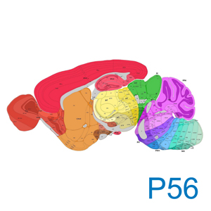
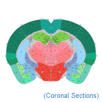
These anatomical reference atlases illustrate the adult mouse brain in coronal and sagittal planes of section. They are the spatial framework for datasets such as in situ hybridization, cell projection maps, and in vitro cell characterization.

3D Viewer
The Allen Brain Explorer (beta) is an application that allows users to browse multimodal datasets in an annotated 3D spatial framework. It is created from a 3D volumetric reference atlas, annotated symmetrically using image data averaged from 1,675 adult mouse brain specimens.
Explore
Coronal Sections
The 2D coronal reference atlas is created from a 3D volumetric reference atlas, annotated symmetrically using an average of data merged from 1,675 brain specimens. It provides spatial context and a common coordinate framework for mapping data in the Allen Mouse Connectivity Atlas, the Allen Cell Types Database (mouse), and the Allen Brain Observatory.

132 virtual planes at 100 µm intervals
Explore
2011 Coronal Sections
The 2D coronal reference atlas is annotated on Nissl sections collected from a single adult male C57Bl6 mouse, postnatal day 56. It provides the spatial context for in situ hybridization-based gene expression in the Allen Mouse Brain Atlas.

132 coronal sections at 100 µm intervals
Explore
2011 Sagittal Sections
The 2D sagittal reference atlas is annotated on Nissl sections collected from a single adult male C57Bl6 mouse, postnatal day 56. It provides the spatial context for in situ hybridization-based gene expression in the Allen Mouse Brain Atlas.

21 sagittal sections at 200 µm intervals
Explore

Adult Human

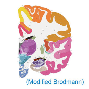
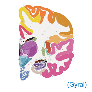
These anatomical reference atlases illustrate the adult human brain, using modified Brodmann or gyral annotation.

Modified Brodmann
The 2D coronal reference atlas is annotated on Nissl sections from a 34-year-old female based on a modified Brodmann nomenclature. It provides spatial context for gene expression in the Allen Human Brain Atlas and the BrainSpan Atlas of the Developing Human Brain.

106 coronal sections at 0.4 - 3.4 mm intervals
Explore
Gyral
The 2D coronal reference atlas is annotated on Nissl sections from a 34-year-old female, by gyrus. It provides spatial context for gene expression in the Allen Human Brain Atlas and the BrainSpan Atlas of the Developing Human Brain.

106 coronal sections at 0.4 - 3.4 mm intervals
Explore

Developing Mouse

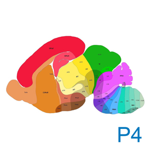
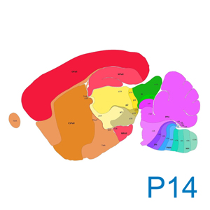
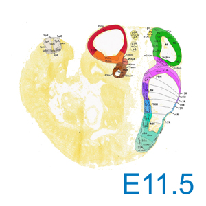
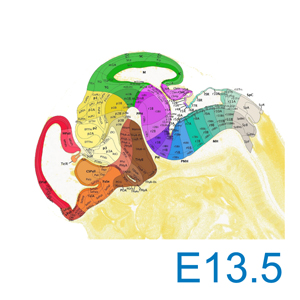
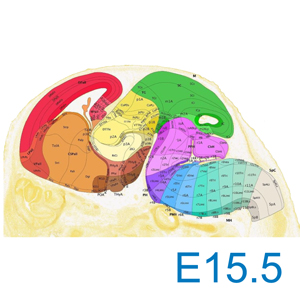
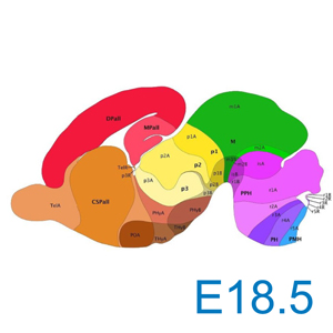
These anatomical reference atlases illustrate the developing mouse brain, covering seven stages of embryonic (E) and postnatal (P) development.

Dr. Luis Puelles used a custom developmental taxonomy for annotation of the Allen Developing Mouse Brain Reference Atlases. Each 2D sagittal reference atlas is annotated on Nissl sections from a single adult made C57B16 mouse.

E11.5
Embryonic day 11.5 (E11.5) or Theiler stage 19 (TS19). 28 sagittal sections at 40 µm intervals.
Explore
E13.5
Embryonic day 13.5 (E13.5) or Theiler stage 21 (TS21). 15 sagittal sections at 100 µm intervals.
Explore
E15.5
Embryonic day 15.5 (E15.5) or Theiler stage 24 (TS24). 16 sagittal sections at 120 µm intervals.
Explore
E18.5
Embryonic day 18.5 (E18.5) or Theiler stage 26 (TS26). 19 sagittal sections at 120 µm intervals.
Explore
P4
Postnatal day 4 (P4). 23 sagittal sections at 160 µm intervals.
Explore
P14
Postnatal day 14 (P14). 39 sagittal sections at 125 µm intervals.
Explore
P56
Postnatal day 56 (P56). 21 sagittal sections at 200 µm intervals.
Explore

Developing Human

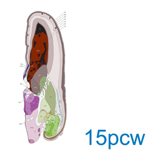
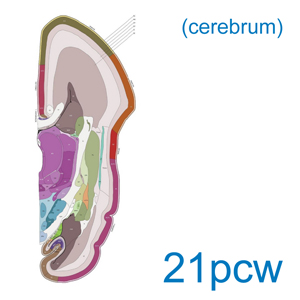
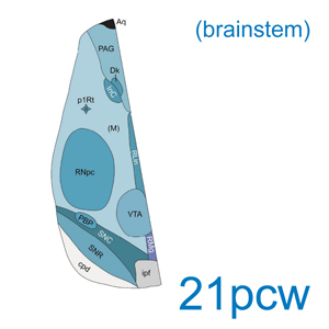
These anatomical reference atlases illustrate the developing human brain, covering two embryonic stages.

15pcw
2D sagittal reference atlas annotated on Nissl sections collected from an embryo at 15 weeks post-conception (15 pcw). 46 coronal sections at 0.5 to 1.0 mm intervals.
Explore
21pcw (cerebrum)
2D sagittal reference atlas annotated on Nissl sections collected from an embryo at 21 weeks post-conception (21 pcw). 81 coronal sections from the cerebrum at 0.5 to 1.2 mm intervals.
Explore
21pcw (brainstem)
2D sagittal reference atlas annotated on Nissl sections collected from an embryo at 21 weeks post-conception (21 pcw). 41 transverse sections from the brainstem at 0.25 to 0.5 mm intervals.
Explore

Mouse Spinal Cord

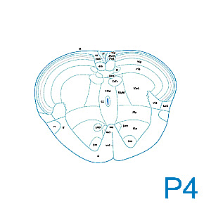
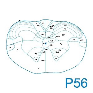
These anatomical reference atlases illustrate the mouse spinal cord in adult and juvenile C57BL/6J mouse. They provide a spatial map for the Allen Mouse Spinal Cord Atlases of gene expression. Dr. Charles Watson and Dr, Gulgun Kayalioglu created a custom taxonomy for annotation of the spinal cord, covering cervical, thoracic, lumbar, sacral, and coccygeal segments.

P4
2D sagittal reference atlas annotated on Nissl sections collected from juvenile male C57BL/6J  mouse, postnatal day 4 (P4). 34 transverse sections at 2 mm intervals.

Note: Image viewer functionality may not be supported in current web browsers
Explore
P56
2D sagittal reference atlas annotated on Nissl sections collected from adult male C57BL/6J  mouse, postnatal day 56 (P56). 34 transverse sections at 2 mm intervals.

Note: Image viewer functionality may not be supported in current web browsers
Explore