Visual Coding

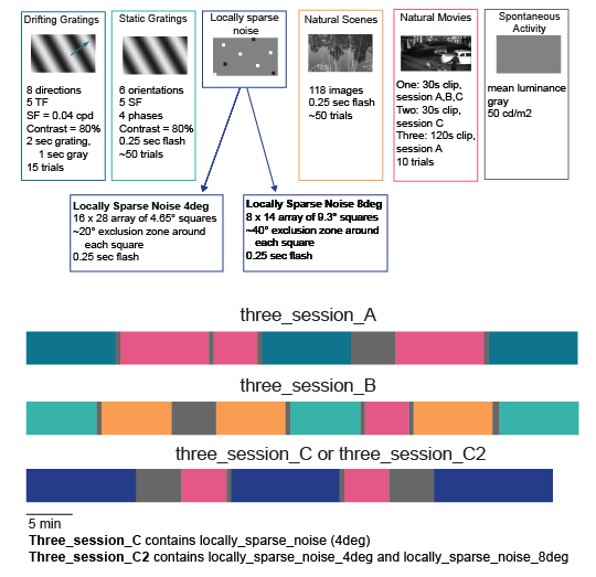
Using a broad range of visual stimuli, ranging from gratings and noise stimuli to natural images and movies, we have surveyed the spatial and temporal dynamics of visual representation in the mouse corticothalamic visual pathways.
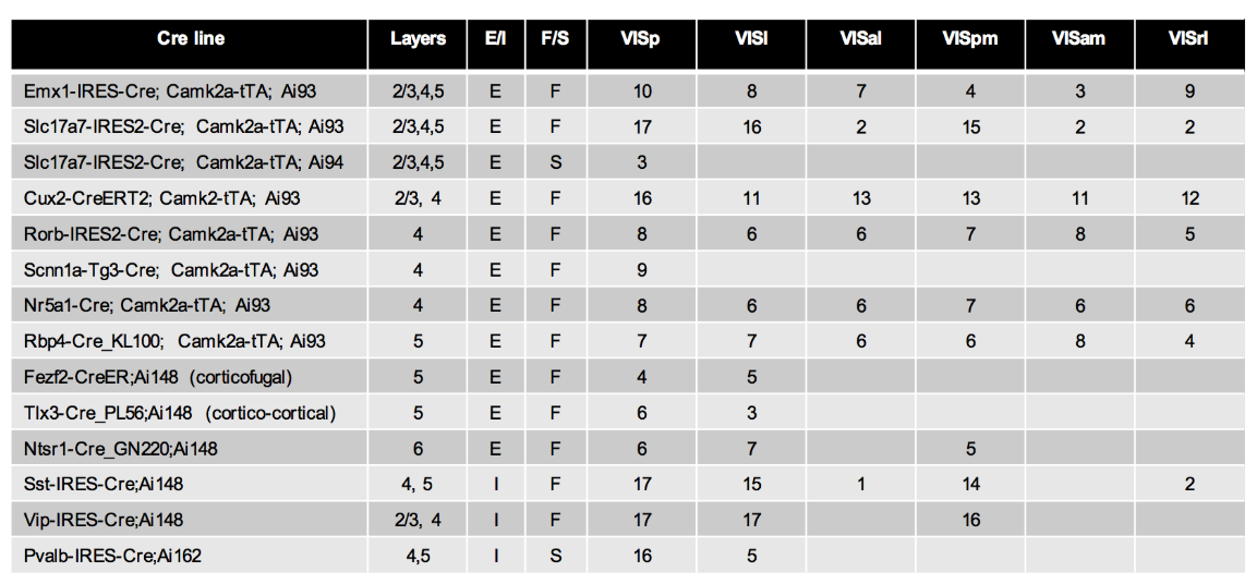
2P Ca+ Imaging

Using transgenically expressed GCaMP6 restricted to specific populations of neurons, we have recorded the visual responses from over 63,000 neurons from 14 transgenic lines, across 6 cortical areas and 4 cortical layers. The spatial and temporal dynamics of the visual responses are measured using a diverse set of visual stimuli. Calcium imaging enables large populations of hundreds of neurons to be imaged simultaneously, allowing the interactions of neurons to be explored.
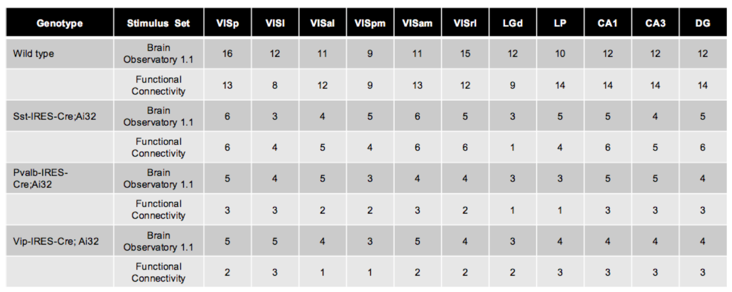
Neuropixels

Using state-of-the-art implantable probes, called Neuropixels, we have recorded the spiking activity of nearly 100,000 neurons from wild-type mice and 3 transgenic lines, across a variety of regions in the cortex, hippocampus, and thalamus. We use a similar set of stimuli to the 2P Imaging Observatory to facilitate a direct comparison between these two datasets. A key feature of these experiments is our ability to record simultaneously across as many as 8 visual regions, which will enable scientists to study inter-areal neural communication patterns in greater detail than ever before.

Visual Coding - 2P Ca+ Imaging

Using transgenically expressed GCaMP6 restricted to specific populations of neurons, we have sampled responses from 14 transgenic lines, across 6 cortical areas and 4 cortical layers. In total 63,251 neurons have been imaged.

Cre lines include

  • Pan-excitatory: Emx1-IRES-Cre, Slc17a7-IRES2-Cre
  • Specific excitatory populations: Cux2-CreERT2 ; Rorb-IRES2-Cre; Scnn1a-Tg3-Cre;Nr5a1-Cre; Rbp4-Cre; Tlx3-Cre; Fezf-CreER; Ntsr1-Cre
  • Inhibitory interneurons: Vip-IRES-Cre; Sst-IRES-Cre; Pvalb-IRES-Cre

More information on Cre lines used in this dataset is available here

Data was collected from primary visual cortex (VISp) and five higher visual areas (VISl, VISal, VISpm, VISam, VISrl).

Visual stimuli

Diverse but well-studied visual stimuli were presented to awake mice in a passive viewing condition. Stimuli included many traditional stimuli such as gratings and locally sparse noise to characterize traditional tuning curves and receptive field properties, as well at natural scenes and movies that have more diverse features and natural statistics. The stimuli were presented in three one-hour imaging sessions per population of neurons.

Watch Team Talk About Analysis

Neuropixels Dataset

Neuropixels represent a major leap forward in neural recording technology. By leveraging CMOS manufacturing, the process that produces the chips inside our cameras and phones, Neuropixels dramatically reduce the size and cost of devices for reading out activity from the brain. Originally developed by imec in collaboration with funding partners at the Allen Institute, the Wellcome Trust, the Gatsby Foundation, and HHMI, Neuropixels are now publicly available via neuropixels.org. Each probe contains 960 recording sites distributed along a shank that's 10 mm long but only 70 microns wide. A subset of 374 or 383 channels can be read out at a time, detecting action potentials from hundreds of neurons with a single probe. Their long shank and compact size makes it possible to insert multiple probes into the same brain, allowing us to study real-time interactions between areas with an unprecedented level of detail.

Target Areas

In our Neuropixels experiments, the same set of cortical visual areas are targeted as in the 2-Photon Imaging Brain Observatory, but all of them can often be recorded simultaneously. Because each probe is 10 mm long, they can extend down to subcortical visual areas, such as lateral geniculate nucleus (LGN) and lateral posterior (LP) nucleus, capturing activity from cortex and thalamus in the same experiment. Spikes in the hippocampal formation (CA1, CA3, and dentate gyrus) are also measured routinely.

In each experiment, mice from one of four different genotypes are used:

  • wild type
  • Sst-IRES-Cre; Ai32 (ChR2)
  • Pvalb-IRES-Cre; Ai32 (ChR2)
  • Vip-IRES-Cre; Ai32 (ChR2

The three transgenic lines allow inhibitory interneuron sub-classes to be identified using optotagging techniques.

Visual Stimuli

Each mouse is exposed to a standardized set of visual stimuli, allowing visual responses across many regions to be mapped in parallel. The stimulus set is designed to help scientists characterize the tuning properties of individual neurons, using classic visual stimuli such as Gabor patches, full-field drifting gratings, and moving dots. A set of natural images and movies are also presented, allowing researchers to explore the interactions between visual areas under more ecologically relevant conditions.

The first release of Neuropixels data contains two separate stimulus sets. The first, known as "Brain Observatory 1.1," shares many of the same stimuli as the 2P dataset, facilitating a comparison of visual responses measured by these two complementary recording modalities. The second, known as "Functional Connectivity," includes a subset of stimuli with a high number of repeats, in order to facilitate the study of inter-areal functional interactions.