Human and Mammalian Brain Atlas Release: Basal Ganglia (pre-print)
Human and Mammalian Brain Atlas (HMBA) is a major atlas of the BRAIN Initiative Cell Atlas Network (BICAN) that proposes to establish a comprehensive, highly granular cell atlas in complete adult human, macaque, and marmoset brains that links brain structure, function and cellular architecture.
Cell Types
Cross-species Taxonomy
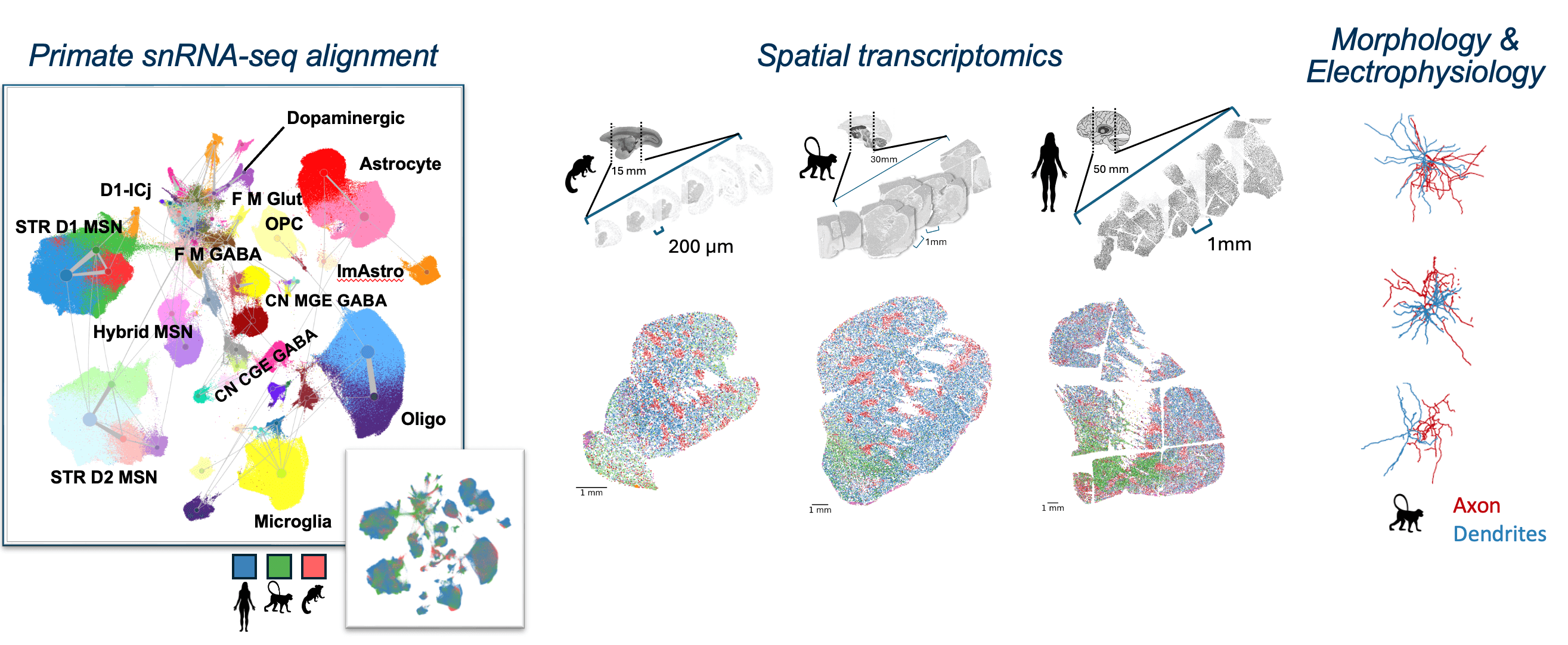
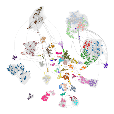
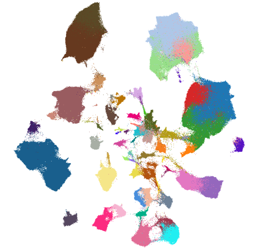
The consensus basal ganglia cell type taxonomy is the result of iterative clustering and cross-species integration of transcriptomic data from single-nucleus 10x Genomics multiomic profiling. The taxonomy encompasses neurons from key structures within the basal ganglia, including the caudate (Ca), putamen (Pu), nucleus accumbens (NAc), the external and internal segments of the globus pallidus (GPe, GPi), ventral pallidus (VeP), subthalamic nucleus (STN), and substantia nigra (SN). By combining data from multiple primate and rodent species, this consensus taxonomy highlights both conserved and species-specific cell types. The taxonomy was validated through marker gene expression analysis, comparison with previously published taxonomies, and self-projection, ensuring the accuracy and robustness of each level in the taxonomic hierarchy.
Data defining the taxonomy
The single-cell transcriptomics data that was clustered to generate this taxonomy is available for visualization, programmatic use, and download.
Integration with Cell Ontology
The consensus basal ganglia cell type taxonomy is incorporated directly into the Cell Ontology (pre-print) - the community standard for cell type annotation used by major single cell data portals such as CZ CELLxGENE and the Broad Single cell Portal. Integration is semi-automated and ensures that the Cell Ontology reflects the taxonomy hierarchy. All HMBA cell types in CL and their markers are linked directly to reference data on the Allen Brain Cell Atlas. All types are linked to locations defined in the Harmonized Ontology of Mammalian Brain Anatomy (HOMBA). Integration into CL also serves to integrate cell types with other standards, including the types defined by the human Cell Atlas and to map types to HMBA defined types to classically defined cell types and their synonyms.
Anatomy
Harmonized Ontology of Mammalian Brain Anatomy (HOMBA)
The Harmonized Ontology of Mammalian Brain Anatomy (HOMBA) is a harmonized cross-species taxonomy of 2341 brain and spinal cord structures. Derived from the Allen Developing Human Brain Atlas (DHBA) ontology, the HOMBA is hierarchical, allowing users to aggregate structures from fine grain parcellations to broad regions. Terminology is harmonized across human, primate, and rodent structures with synonymous terms and includes transient developmental structures. HOMBA is designed for neuroanatomical applications including brain sampling and dissection, tissue block mapping, atlas building, cell-type and pathology localization, and linking cross-species and developmental datasets.
Brain structures and templates in 3D space
Access and use all anatomy assets programmatically
The Common Coordinate Framework Multispecies Atlas Primer (CCF-MAP) is a resource for accessing marmoset, macaque, and human atlases developed by the Human and Mammalian Brain Atlas (HMBA) consortium, as well as the CCFv3 mouse atlas from the Allen Institute. CCF-MAP is a repository of publicly accessible MRI templates, associated parcellations of basal ganglia structures, and related metadata, which can be viewed and downloaded. CCF-MAP also includes Jupyter notebook tutorials for querying voxel data and for downloading region masks for offline use.
Tools
Explore the data: ABC Atlas
The Allen Brain Cell (ABC) Atlas provides a platform for visualizing multiple multimodal single cell data datasets simultaneously. Four Basal Ganglia data sets are now available in the ABC Atlas for exploration. Each data set is annotated with the Mammalian Basal Ganglia Consensus Cell Type Taxonomy. Human, marmoset, and macaque spatial data sets include harmonized ontology of the mammalian brain anatomical annotations.
Map your data to the taxonomy: MapMyCells
From your browser
MapMyCells allows you to assign cell type names from Allen Institute-hosted taxonomies to your own single cell and spatial transcriptomics data. The Mammalian Basal Ganglia Consensus Cell Type Taxonomy is now available as a mappable taxonomy in MapMyCells. Our mapping algorithm now includes species detection and automatic ortholog conversion.
In Python
An example notebook is provided showing how to map unlabeled data onto the taxonomy using the Python version of MapMyCells. At this point, files are separated by species. Human data should be mapped with the human data files, macaque data with the macaque data files, and marmoset data with the marmoset data files.
Human: precomputed_stats in AWS S3 --- query_ markers in AWS S3
Macaque: precomputed_stats in AWS S3 --- query_ markers in AWS S3
Marmoset: precomputed_stats in AWS S3 --- query_ markers in AWS S3
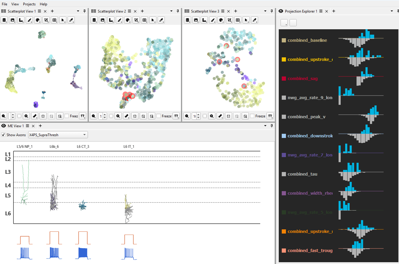
Analyze the atlas visually: Cytosplore Viewer
Cytosplore Viewer provides a complementary data analysis and visualization to ABC Atlas, focusing on fast local compute for the linked exploration of multi-modal spatial and single cell datasets. It offers on high interactivity, low-latency and on-the-fly analyses, complementing the BKP functionality with interactive, visual analytics across multiple datasets, providing for instance:
- Deep dives into single-cell and spatial data for in-depth re-analysis of subsets of cells directly in Cytosplore Viewer. Specific niche functionalities are on-the-fly re-embedding, local gene expression gradients detection, differential expression analysis and manual annotation / correction facilities.
- Comparative visualization for direct comparative analyses of single cell and spatial data across multiple species and / or between brain regions. Central in these comparisons is the identification of conserved and diverging features patterns across these datasets and relate these to evolutionary and anatomical prior knowledge.
- Multimodal single-cell feature integration: for linked visualization of for instance patch-seq data (for exploration of annotated t-typed cells in relation to their morphological and electrophysiological features) and ATAC-seq / spatial / RNA-seq combinations.
Access and analyze atlas programmatically
The Allen Brain Cell (ABC) Atlas Access package provides methods for downloading the HMBA-BG data and metadata programmatically via python. The repository also provides Jupyter notebooks showing examples of how to join and utilize these data products, allowing users to delve deeper into visualizations and results they create with the ABC Atlas visualization tool.

Connectome Workbench for visualizing CCFs and transcriptomic samples
The Connectome Workbench platform (Get Connectome Workbench) enables visualization, navigation, and analysis of human, macaque, and marmoset brain templates, atlases, and canonical coordinate frameworks (CCFs), including volume-based representations of subcortical structures and surface-based representations of cerebral cortex that respect the topology of the cortical sheet.
Connectome Workbench also plays a key role in encoding the precise location of tissue samples used for HMBA single-cell multiomics data and in visualizing spatial transcriptomic data after precise alignment to corresponding MRI slices. Datasets associated with multiomic and spatial transcriptomic data localization will be shared via the BALSA database.
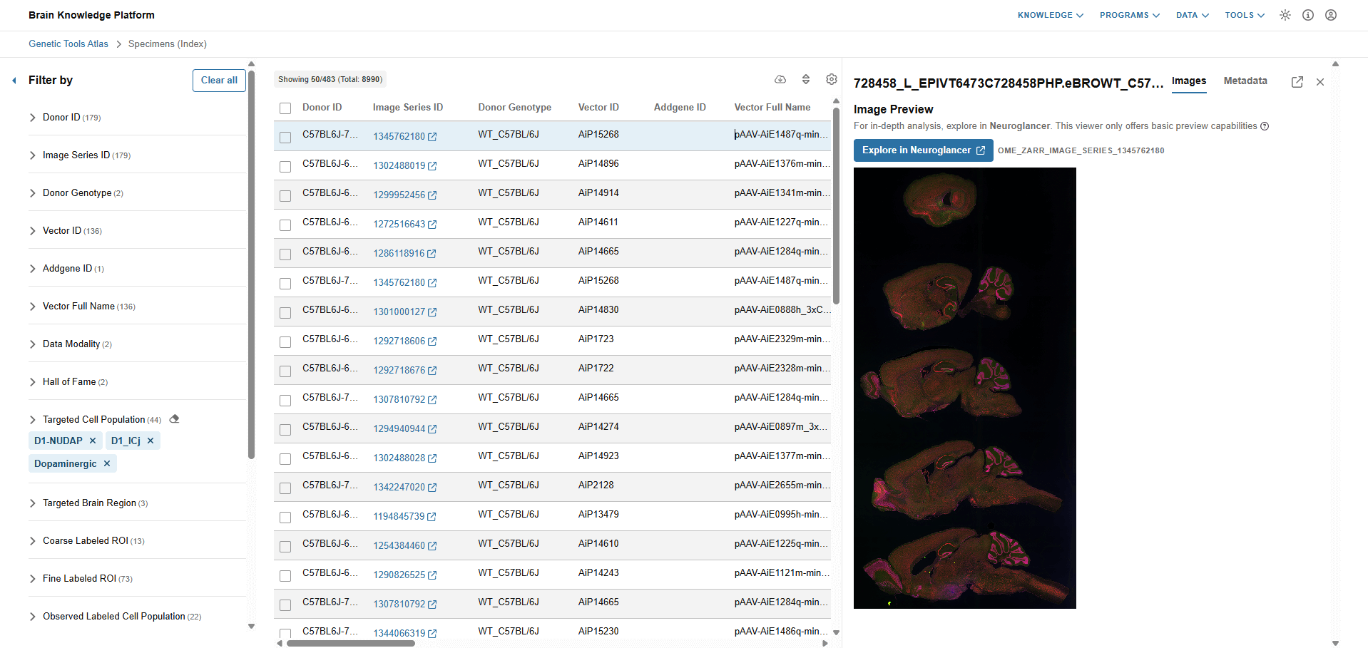
Run your own experiments: Genetic Tools Atlas
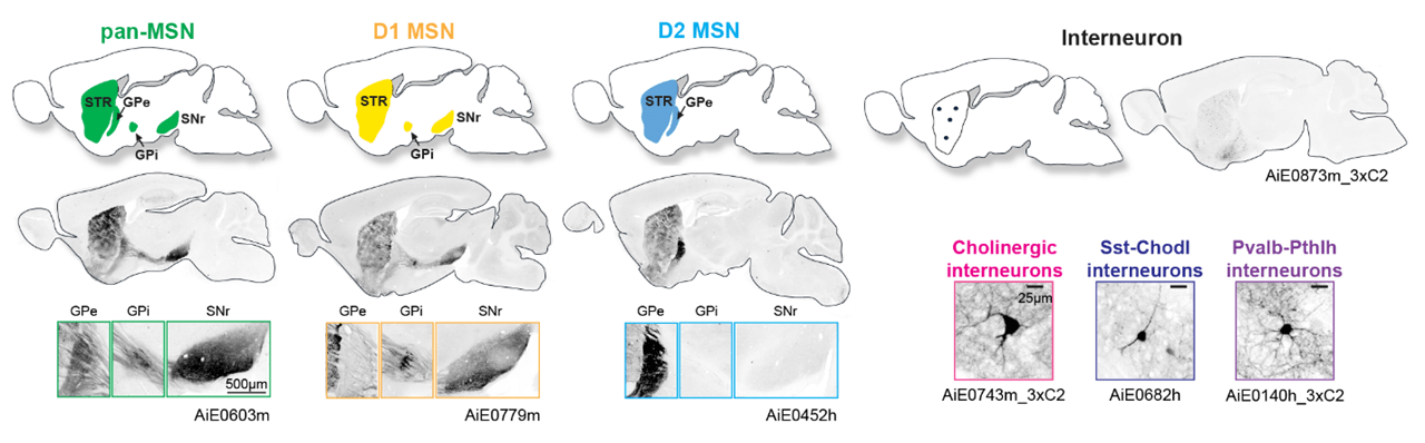
The Genetic Tools Atlas (GTA) is a dynamic, evolving resource for exploring and accessing diverse cell types in the brain. This expansive collection of datasets highlights tools that enable high-precision targeting of cell types and circuits across the basal ganglia in mouse and macaque. Among its highlights are novel tools for labeling granule cells in the Islands of Calleja (OT D1 ICj), non-canonical MSNs such as D1 NUDAP (STRv D1 NUDAP MSN), and midbrain dopaminergic cell populations (see first link below). Further details of tool design are presented in Hunker, Wirthlin, et al. 2024, with most tools available on Addgene (see second link below of a publication centered view of GTA).
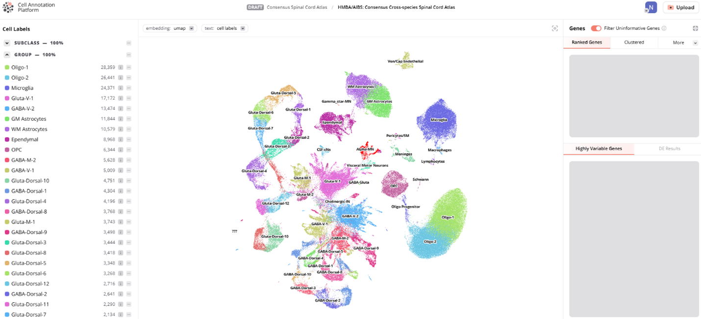
Improve our cell type names: Cell Annotation Platform
The Cell Annotation Platform (CAP) offers a community-driven, centralized location where researchers can easily manage and share their cell annotations for single-cell RNA-sequencing datasets. Widely used by cell typing communities in the Human Cell Atlas, CAP provides a platform for users of the Mammalian Basal Ganglia Consensus Cell Type Taxonomy to provide input on cell type names and to add context. Get started by following the instructions on our Annotation Jamboree website (note that the “Spatial transcriptomic cirrocumulus” visualization shave been moved to the ABC Atlas).
Data
Data licensing information for these data resources can be found in Brain Knowledge Platform's Data Catalog.
Single-cell and spatial transcriptomics
ATAC-seq
ATAC-seq data are available at various stages of processing with snaptac2: (1) fragment files in .h5ad format, (2) Group level bigwigs and (3) MACS3 peak calling. Data for all species and levels of processing can be found at the public AWS s3 bucket: s3://hmba-bican-sharing-802451596237-us-west-2/BasalGanglia_pre-print_ATAC/.
Patch-seq
This release also includes more than 1,000 patch-seq cells, sampled from across the Basal Ganglia in macaque. Multimodal data files, cell features, and cell type labels from the consensus taxonomy will soon be available for download. Data can be visualized in the Cytosplore Viewer as well. (See Tools section above)
Methods
Consensus cell type nomenclature
To unify the field’s understanding of basal ganglia cell types across species, efforts supported by the NIH BRAIN Initiative have initiated a comprehensive nomenclature system that synthesizes pre-existing literature describing cell type nomenclature with new cross-species transcriptomic data. This effort includes coordinated contributions from the Human and Mammalian Brain Atlas (HMBA) project in the BRAIN Initiative Cell Atlas Network (BICAN), and the Basal Ganglia AAV Toolbox project of the BRAIN Initiative Armamentarium for Precision Cell Access. This initiative integrates HMBA single-nucleus RNA sequencing (snRNA-seq) data from human, macaque, marmoset, and previously published mouse basal ganglia, with the goal of generating a consensus cell type taxonomy that can be widely adopted by the scientific community. By focusing on conserved marker genes and shared molecular profiles to supplement established names from the broader community, we have developed a standardized naming system that captures the evolutionary relationships and functional distinctions among basal ganglia cell types. The HMBA consensus basal ganglia taxonomy is designed to streamline communication, foster collaboration, and facilitate the development of novel research tools targeting specific cell types across multiple species.
Cell type taxonomy
Our consensus basal ganglia cell type taxonomy is the result of iterative clustering and cross-species integration of transcriptomic data from single-nucleus 10x Genomics multiomic profiling. The taxonomy encompasses neurons from key structures within the basal ganglia, including the caudate (Ca), putamen (Pu), nucleus accumbens (NAc), the external and internal segments of the globus pallidus (GPe, GPi), ventral pallidus (VeP), subthalamic nucleus (STN), and substantia nigra (SN). By combining data from multiple primate and rodent species, we have developed a consensus taxonomy that highlights both conserved and species-specific cell types. We validate our taxonomy through marker gene expression analysis, comparison with previously published taxonomies, and self-projection, ensuring the accuracy and robustness of each level in the taxonomic hierarchy.
Sample and cell type metadata collection
To support the HMBA consensus cell type taxonomy, we have compiled extensive metadata providing detailed information associated with each identified cell type. These metadata include information on gene expression patterns and marker genes from snRNA-seq as well as synonymous names from existing literature, including Yao et al. 2023 and Siletti et al 2023. The metadata fields will be organized under an aligned taxonomy data format enabling integration with pre-existing tools such as CellxGene and Cell Annotation Platform.
Design and build genetic tools (enhancer AAVs)
In collaboration with the BRAIN Armamentarium Consortium, we have developed an extensive collection of cell type-specific viral tools, designed to target and manipulate specific cell types identified in our cross-species integrated consensus BG taxonomy (Hunker, Wirthlin, et al. 2024). Using bulk or single nucleus ATAC-seq and multiomic data, we identify cell type-specific gene regulatory elements—particularly enhancers—linked to the cell types we identified through snRNA-seq. These efforts have culminated in a robust computational pipeline for identifying successful enhancers with a high degree of accuracy, the Cross-species Enhancer Ranking Pipeline (CERP). This pipeline allows us to identify and prioritize testing of the most promising candidate cell type enhancers for targeting homologous cell types across mammalian species. This viral tool collection comprises hundreds of enhancer adeno-associated virus (AAV) vectors, each with detailed brain expression analysis catalogues in the Genetic Tools Atlas. The most promising tools were also subjected to molecular validation. Additionally, we offer variants of these tools with enhancers optimized for stronger or more specific labeling, as well as variants delivering fluorescent proteins, channelrhodopsin variants, GCaMP variants, recombinases (Cre and Flp) to enable targeted experimental manipulation for functional studies. Many of these enhancer AAV vectors are already available through Addgene, with new tools continuously being added to the collection. These collective resources provide researchers with a powerful toolkit for targeting and studying the diverse cell types of the basal ganglia across multiple species to advance our understanding of BG function and dysfunction in health and disease.
Cross-species Enhancer Ranking Pipeline (CERP)
Cell type subclasses in BG regions are conserved between mouse, human, and non-human primates. Candidate enhancers were identified for the major BG cell subclasses using a novel computational pipeline we call Cross-species Enhancer Ranking Pipeline (CERP), which examines open chromatin using single nucleus ATAC-seq datasets. Additional enhancers were selected using CERP to target other BG cell populations, including groups of cell types with regional enrichment, such as dorsal vs. ventral striatum.
Acknowledgments
Supported by the NIH BRAIN Initiative Armamentarium grant UF1MH128339 and Cell Atlas Network grant UM1MH130981.