Our Science (SEA-AD)
The Seattle Alzheimer’s Disease Brain Cell Atlas (SEA-AD) consortium aims to provide scientific insight on the pathogenesis of Alzheimer’s disease using data generated by SEA-AD and others. Access our latest published work describing cellular and molecular changes in AD progression.
Note: Code for reproducing analyses from some of these papers is now available via the "Our Code" landing page.

Platform papers
Large studies from the whole SEA-AD consortium describing major data generation efforts and associated knowledge from these data sets.

Multiregional single-cell profiling reveals shared andspecialized cellular vulnerability in Alzheimer’s disease (Travaglini,Gabitto, Ding, et al., bioRxiv, 2026)
This paper extends the SEA-AD atlas across ten neo- and allocortical brain regions to determine whether the cellular changes associated with Alzheimer’s disease are shared across the brain. By analyzing the molecular profiles of roughly seven million cells and relating them to regional pathology, the researchers found that vulnerability is remarkably consistent across regions, with the same subsets of inhibitory neurons, oligodendrocytes, and glia changing in a stereotyped sequence that begins before symptoms emerge. The study also uncovered unexpected vulnerability in specialized cell populations, including primary visual cortex layer 4 excitatory neurons previously thought to be resilient, and identified neuronal hyperexcitability as a potential common mechanism linking both early- and late-vulnerable cell types.
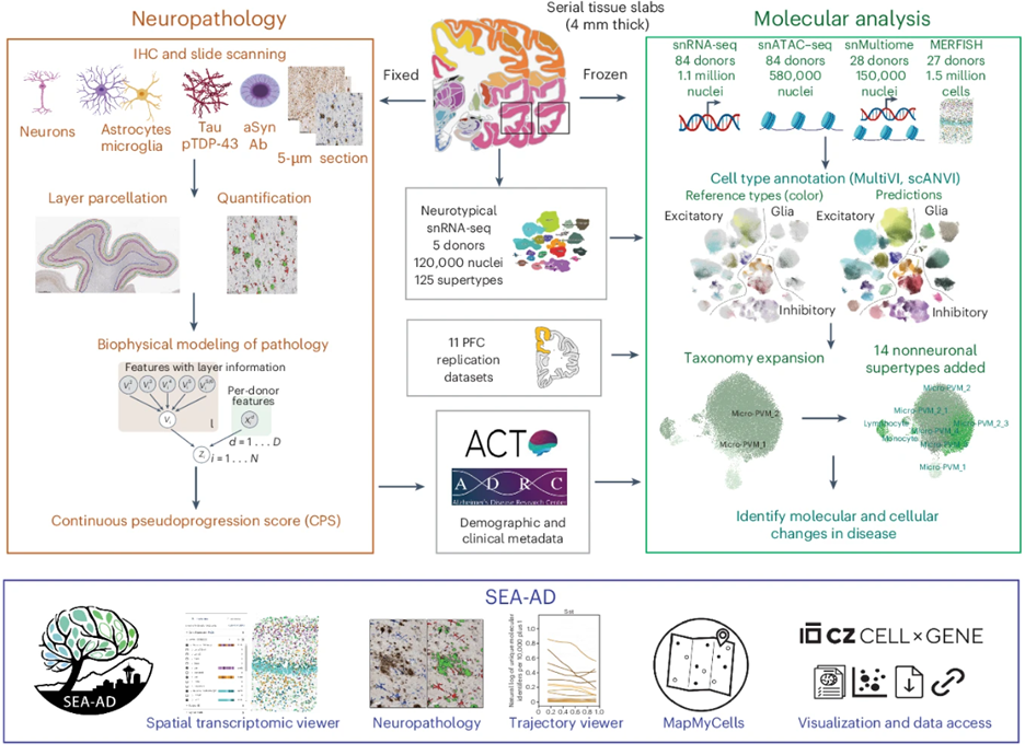
Integrated multimodal cell atlas of Alzheimer’s disease (Gabitto, Travaglini, et al., Nature Neuroscience, 2024)
This paper describes the creation of a massive molecular and cellular atlas of middle temporal gyrus to better frame and understand how Alzheimer’s disease progresses. By analyzing the genetic activity of millions of individual cells, the researchers discovered that the disease actually unfolds in two distinct phases: an early, slow-moving stage where specific Sst+ interneurons are lost and inflammation begins, followed by a later stage of accelerated neuropathology and loss of excitatory neurons and Pvalb+ and Vip+ inhibitory neuron subtypes. These findings were replicated in prefrontal cortex in SEA-AD and other major AD studies.
Focused studies
Other papers with SEA-AD researchers as primary authors focused on SEA-AD data sets or novel computational algorithms.
The Caudate Nucleus Exhibits Distinct Pathology and Cell Type-Specific Responses Across Alzheimer’s Disease (Kana et al., bioRxiv, 2026)
This paper represents the characterization of the Alzheimer's pathological spectrum within the caudate nucleus. Utilizing digital pathology, researchers were able to confirm a less pronounced phosphorylated tau and amyloid-β burden when compared to other cortical areas. Phosphorylated tau and amyloid-β were also shown to localize separately into white and gray matter, respectively. Both, in part, explain the lack of neuronal cell death observed in the caudate even in high burden parts of the spectrum. However, researchers were able to identify glimmers of a global phosphorylated tau response in microglia.
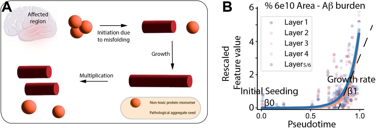
B-BIND: Biophysical Bayesian Inference For Neurodegenerative Dynamics (Agrawal et al., Annals of Applied Statistics, 2026)
This paper introduces B-BIND, a new mathematical framework designed to track how Alzheimer’s disease progresses by analyzing the buildup of various pathological proteins. Because researchers often only have "snapshots" of the brain from different individuals, this model uses a "pseudotime" scale to rank patients along a continuous timeline of disease severity. Ultimately, this framework lays the groundwork for identifying the hidden biological stages of neurodegeneration and the specific cellular changes that drive the disease over time.
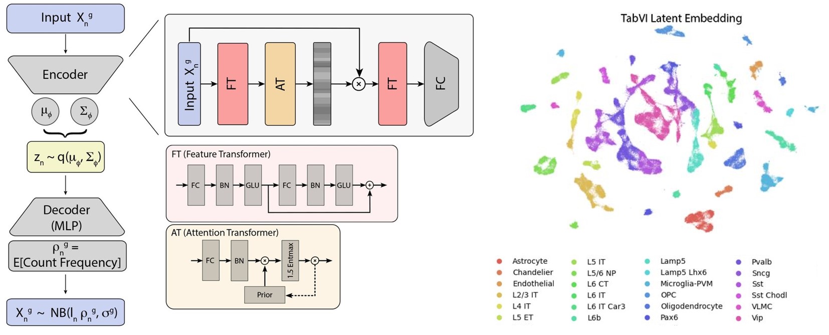
TabVI: Leveraging Lightweight Transformer Architectures to Learn Biologically Meaningful Cellular Representations (Chandrashekar et al., bioRxiv, 2026)
This paper introduces TabVI, a lightweight AI model designed to analyze single-cell genomic data more effectively than standard language-based AI. By adapting "transformer" technology—the same tech behind ChatGPT—to better fit the non-sequential, hierarchical nature of genes, TabVI can more accurately identify cell types and biological patterns. This tool provides a more efficient and interpretable way for researchers to study how diseases like Alzheimer’s affect individual cells across large datasets.
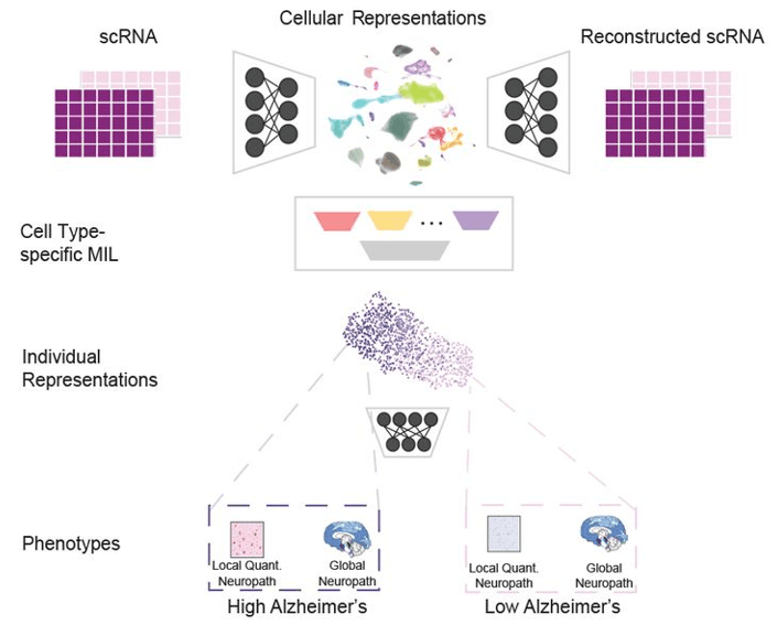
scVIP: personalized modeling of single-cell transcriptomes for developmental and disease phenotypes (Lai et al., bioRxiv, 2026)
This paper introduces scVIP, an AI framework designed to reconstruct how Alzheimer’s disease progresses by combining molecular measurements from individual cells with information about each donor’s disease severity. By learning personalized disease trajectories, scVIP can more accurately predict neuropathology, identify the cell types most affected by disease, and connect findings across independent studies. This approach provides researchers with a powerful new tool for exploring the cellular and molecular changes that drive neurodegeneration.
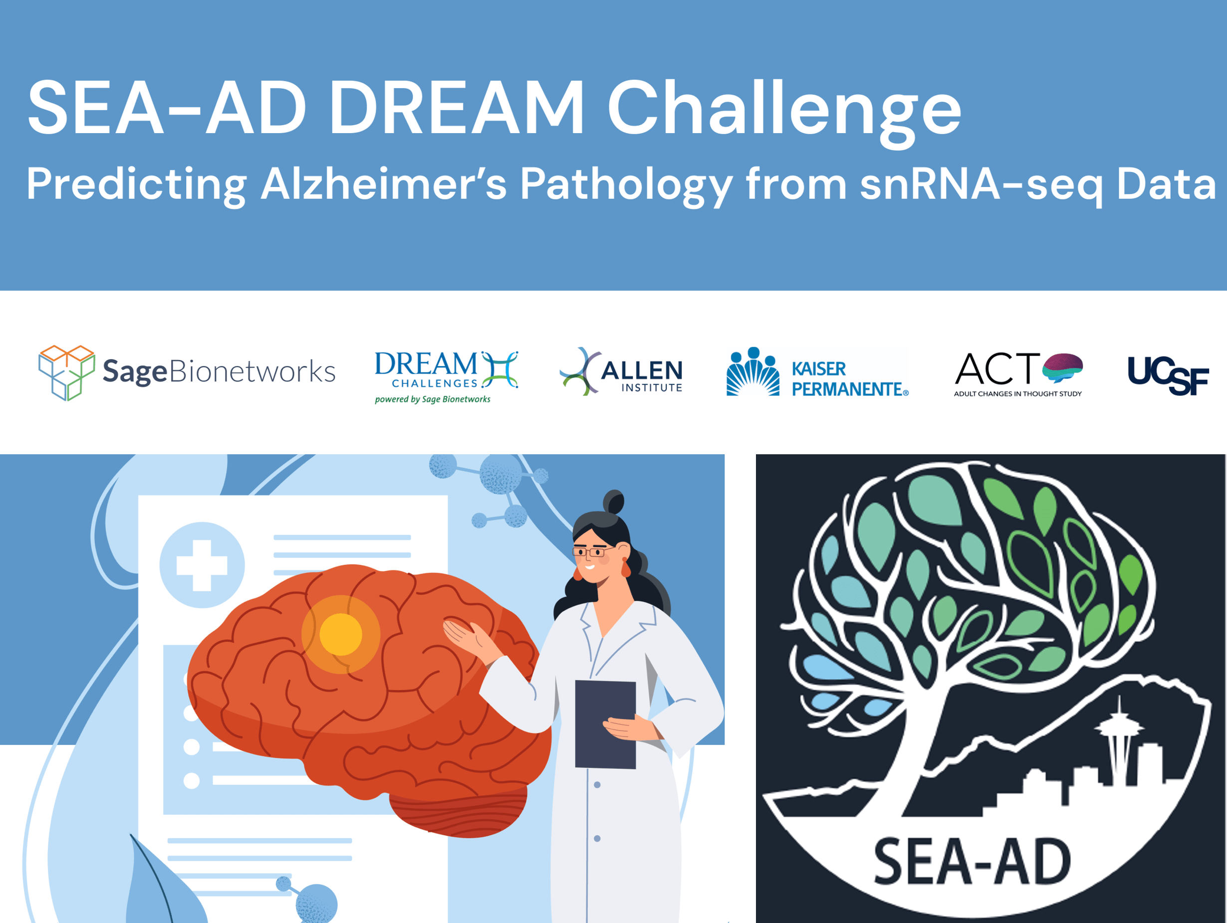
The SEA-AD DREAM Challenge: Community benchmarking human and AI agent solutions for Alzheimer's disease neuropathology prediction from single-nucleus transcriptomics (Lai et al., bioRxiv, 2026)
This paper presents the SEA-AD DREAM Challenge, an open international competition in which 17 teams from 15 countries built models to predict Alzheimer's disease neuropathology from single-nucleus RNA-sequencing data. The best models predicted categorical disease staging with near-perfect accuracy and estimated amyloid-β and tau burden competitively, and the challenge also debuted the first AI Agent Track, establishing a reproducible benchmark for molecular neuropathology prediction.
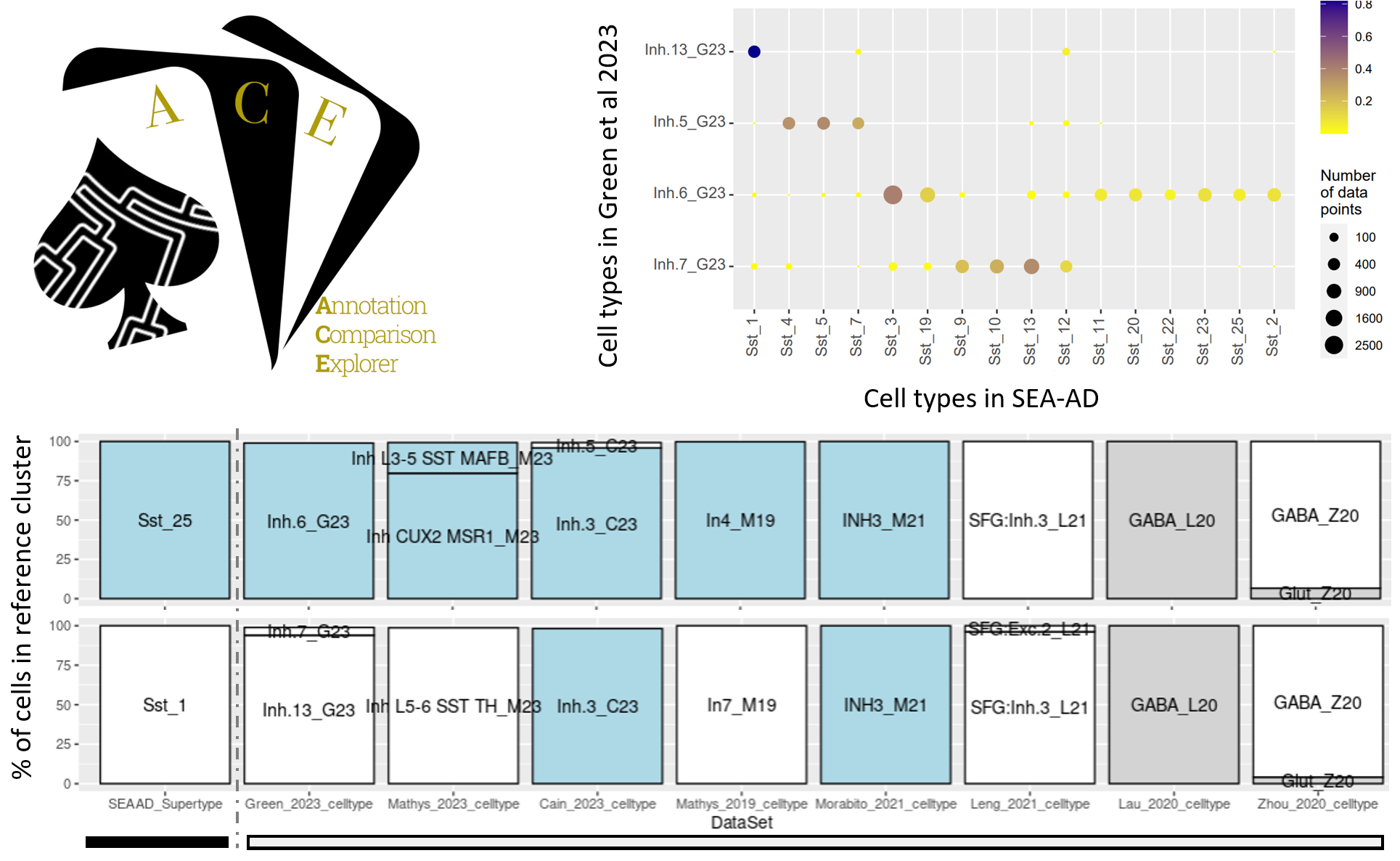
Annotation Comparison Explorer (ACE): connecting brain cell types across studies of health and Alzheimer’s Disease (Miller et al., bioRxiv, 2025)
This paper introduces Annotation Comparison Explorer (ACE), a web-based tool developed by the Allen Institute to address the challenge of comparing cell-type classifications across different brain studies. ACE allows researchers to map their own single-cell data to established taxonomies, such as the Seattle Alzheimer’s Disease Brain Cell Atlas (SEA-AD), and compare annotations like donor demographics and disease-related changes. By applying ACE to multiple published Alzheimer's datasets, the authors identified consistent signatures of the disease, such as a decrease in specific somatostatin interneurons, demonstrating the tool's utility in unifying diverse datasets to advance the understanding of brain health and neurodegeneration.
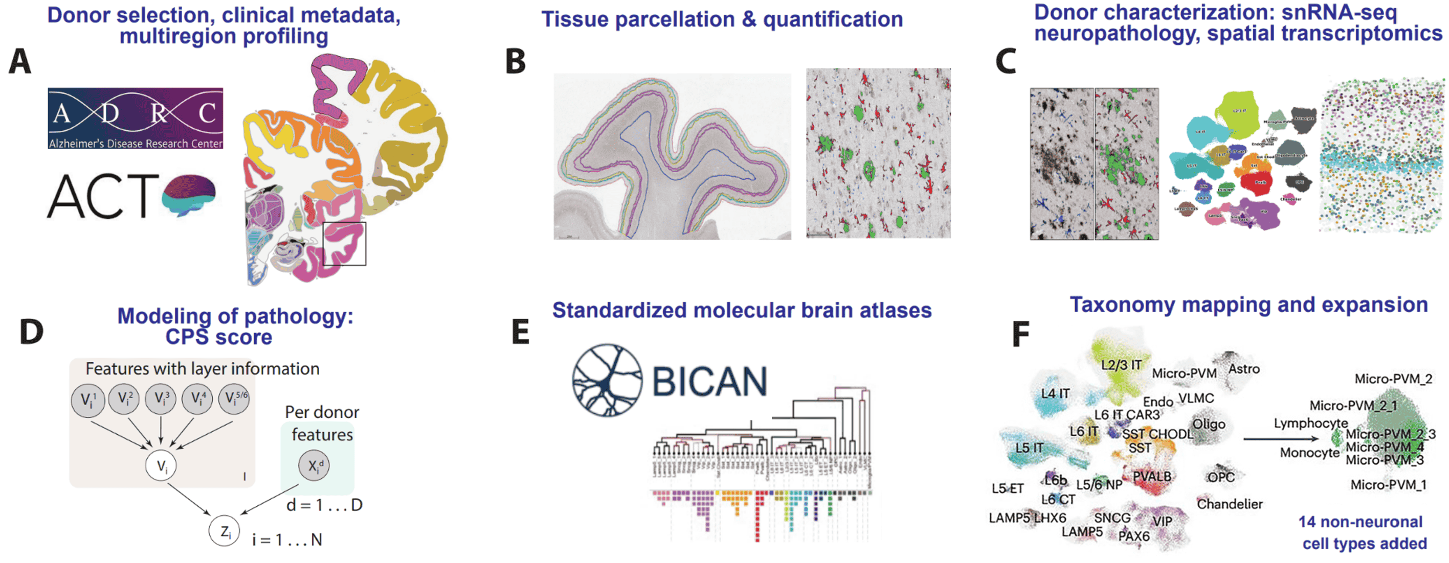
SEA-AD is a multimodal cellular atlas and resource for Alzheimer’s disease (Hawrylycz et al., Nature Aging, 2024)
This commentary on Gabitto, Travaglini, et al 2024 presents SEA-AD as a multifaceted open community resource designed to identify cellular and molecular pathologies that underlie Alzheimer’s disease. Integrating neuropathology, single cell and spatial genomics, and longitudinal clinical metadata, SEA-AD is a unique resource for studying the pathogenesis of Alzheimer’s and related dementias.
Collaboratory studies
Projects led by other folks in the AD community, but with significant SEA-AD input or using SEA-AD data resulting in co-authorship by SEA-AD researchers.
Somatic cancer variants enriched in Alzheimer’s disease microglia-like cells drive inflammatory and proliferative states (Huang et al., Cell, 2026)
Using transcriptomic datasets (including SEA-AD), this study reveals that somatic mutations—genetic changes occurring after birth—are twice as common in Alzheimer’s brains than in healthy, age-matched controls. These mutations are concentrated in the brain's immune cells (microglia) and often affect "cancer driver" genes, causing the cells to multiply and trigger inflammation. This suggests that Alzheimer's may be driven by a tumor-like process where mutated immune cells expand throughout the brain and accelerate neurodegeneration.
Read the paper | Use RNA-MosiacHunter
Single Cell Landscape of Sex-specific Drivers of Alzheimer’s Disease (Wu et al., Alzheimer’s & Dementia, 2025)
This study analyzed single nucleus transcriptomics data (including from SEA-AD) to understand how Alzheimer's disease affects men and women differently at a genetic level. Researchers found female-specific gene associations providing "protective" effects in neurons of dorsolateral prefrontal cortex, while specific inflammatory signals in microglia may increase risk for protein buildup in women. These findings highlight that the molecular path to cognitive decline is sex-specific, pointing toward new, targeted genes for future treatment research.
Read the paper | Access the SEA-AD data | Access the ROS/Map data
CRISPR screens in iPSC-derived neurons reveal principles of tau proteostasis (Samelson et al., Cell, 2026)
This study utilizes genome-wide CRISPRi screens in human iPSC-derived neurons to identify cellular factors regulating the accumulation of tau aggregates, a primary driver of Alzheimer’s disease. The researchers identified the ubiquitin ligase CUL5-SOCS4, alongside pathways such as UFMylation and mitochondrial function, as critical modifiers of tau stability and potential therapeutic targets. Furthermore, the team leveraged SEA-AD data to validate these findings, demonstrating that these tau-regulating genes exhibit altered expression within the specific cell types most vulnerable to Alzheimer’s in human donors.
Read the paper | Explore CRISPR resources
Studies citing SEA-AD
Recent studies citing SEA-AD publications or data, illustrating SEA-AD's research community impact
Title
Author
Year
Source
Type
Abdel-Haleem et al
2026
Brain
Journal article
Almeida et al
2026
Neural Regeneration Research
Journal article
Chaubey et al
2026
Cell Reports Medicine
Journal article
Chu et al
2026
Free Radical Biology and Medicine
Journal article
Dalley et al
2026
Preprint
Dharshini et al
2026
Nature Communications
Journal article
Duggan et al
2026
Molecular Neurodegeneration
Journal article


Explore our Programs
The Allen Institute Institute for Brain Science is active in a wide variety of areas to accelerate progress towards understanding the brain. Find choice resources below.